 APcis ...creative CRO
In obese murine models, CT imaging method for follow up studies of adipose tissue distribution and quantification of visceral and subcutaneous fat are still lacking. Current small animal micro-CT involves long-term X-ray exposure precluding longitudinal studies.
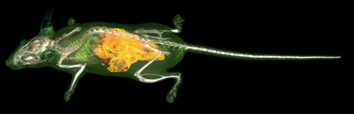
We have overcome this limitation by using a human medical CT which allows very fast 3D imaging (2 sec) and minimal radiation exposure. This work presents novel methods fitted to in vivo investigations of mice model of obesity, allowing automated detection of adipose tissue in abdominal regions of interest and quantification of visceral and subcutaneous fat.

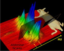
For each mouse, 1000 slices (100µm thickness, 160 µm resolution) were acquired in 2 sec using a Toshiba medical CT (135 kV, 400mAs). A Gaussian mixture model of the Hounsfield curve of 2D slices was computed with the Expectation Maximization algorithm.
Our results show that medical CT imaging combined with automatic image analysis provide precise and reproducible quantification of adipose tissue in mice in vivo allowing repetitive examinations for longitudinal studies.